PIXE – Particle-Induced X-ray Emission
PIXE – Particle-Induced X-ray Emission
PIXE is a powerful method for multi-elemental analysis of solid samples at low concentrations. Without destroying the material, PIXE gives a discrete spectra of X-ray emissions characteristic of the atoms included in the sample, and their different concentrations. PIXE is a highly advanced technique that uses a particle accelerator to expose the sample with a proton beam.
We at Emmace take care of the sample preparation from your device/inhaler, the measurement, as well as the evaluation and explanation of data together with experienced PIXE experts, which are located at Lund University.
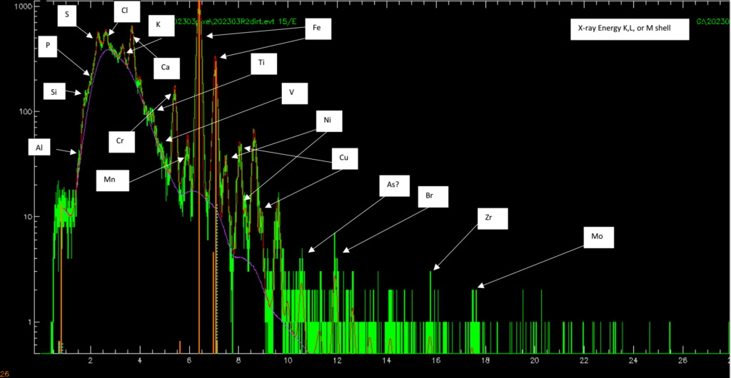
Figure 1. Example of PIXE spectrogram. Each white frame indicates different elements.
PIXE can be used in a variety of applications. One application is to do elemental analysis of aerosol particles emitted from a medical device or inhaler. Another application is to detect trace elements in biomedical samples.
PIXE irradiates the material with a high-energy proton beam (He2+ or even heavier ions can also be used). The high energy of the ions excites the atoms by removing core electrons. The vacancies are filled with electrons from outer shells. When the atom is de-excited an electromagnetic wave is sent out with the same energy as that of the energy gap between the electron shells. The gaps between electron shells are characteristic for each type of atom.
From the X-ray energy spectrum, it is thus possible to deduce the material content of the specimen and the concentration of each element.
An advantage of PIXE compared to electron-beam induced methods, is that the background signal is lower in relation to the measuring signal. This enables detection of low concentrations.